Etikettarkiv: bioimaging

Visual studies and the body

From Sade Kahra’s installation Wear Your Fear – Cancer Collection at BioCity Turku. Copyright Sade Kahra 2018. Photo: Fred Andersson.

Visual studies belongs primarily to the ”humanistic disciplines” or ”the Arts”. To be active in Visual studies has often been a way to combine Art history with Comparative literature, Media or Film studies, Sociology, and even Linguistics and Psychology. However, the development of Image and Media technology takes place in the fields of Science, the fields of numbers and precise instruments. At least some knowledge of the natural sciences and of scientific methods is helpful if you want to understand the use and impact of images today. With our Visual studies intitiative at Åbo akademi, we try to break the comfort zone of our ”building” (Humanities and Languages) and to create dialogues with researchers from Computer Science, Psychology/Medicine, Bioscience and other natural sciences. One opportunity for such dialogues is our course Visuality and Visualization of Information (see Våra kurser / Our Courses).

Our role as ”humanists” is of course not to try to become matematicians or biologists, but to understand (critically) how Science alters our ideas and capabilities. Not the least when it comes to our bodies. Here in Turku, we have a lot to learn from the Bioimaging program at BioCity Turku. Recently, on August 23rd – 24th, BioCity arranged the international symposium Seeing the Invisible. Many artists, working with ”fine arts”, take inspiration from biotechnology and bioimaging. Part of our job as scholars of ”liberal arts” (history, criticism, and the like) is to comment upon their work.

Detail from Sade Kahra’s installation Wear Your Fear – Cancer Collection at BioCity Turku. Copyright Sade Kahra 2018. Photo: Fred Andersson.

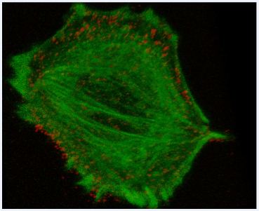
For the Seeing the Invisible symposium, Turku artist Sade Kahra had installed textile objects from her project Wear your Fear – Cancer Collection in the BioCity entrance hall. The objects are white robes on which colourful images of cancer cells have been printed. These images have been produced by scientists who work with the advanced microscopy and visualization devices available at the Bioimaging program. The robes are held together by ribbons on which some background information about each case/image is printed, the information being only partly visible. Most robes are hanging suspended around one of the main staircases of the hall. This exhibition is on view for a month, until September 28th.

Until one realizes that the images printed on the robes actually represent cancer cells, they may only strike the viewer as exceedingly beautiful. Biomedical visualization techniques produce images which are aesthetically very appealing, which makes them increasingly popular and useful for creating curiosity. However there is a difference between curiosity and understanding. In the text which she has written for the exhibition Sade Kahra implies that most of us still interpret scientific images according to old expectations and conventions, whilst it is much harder to fathom the new possibilities and discoveries which these pictures actually make visible:

”Bioimaging is not about reproducing, but depicting reality with a specific meaning. For centuries our eye has been trained to look at images based on certain rules, with an image perception typical of our culture. Common man interprets and perceives the biomedical visualizations as traditional images and therefore, despite their testimonial value, they remain a composition of colors and abstraction.”

To print the images on cloth and to wear them on our bodies could be a way to reclaim the bodily and aesthetic experience in the abstract world of science: ”New technologies have formed medicine into an image science, but as in early physics, our internal microscopic elements are disconnected from all bodily and social contexts.” If this is so, then a more intimate relationship with Science and a deeper understanding of its images may help us identify more clearly the cultural habits and rules which usually condition how we see things. This is no doubt an important aim of Visual studies.

Understanding Basic Vision

Contemplation of Basic Vision, according to the famous philosopher Descartes (1637)

Contemplation of Basic Vision, according to the famous philosopher Descartes (1637)

This is a little report on what we have been doing the past weeks in the Visual studies program. We now start every academic year in English, and with perception. As the title of our course book Basic Vision says, the perception course is really about the very basics of vision and brain. ”Why do you need vision science in visual studies?” some people ask, believing that visual studies is not a scientific field.

They are partly right, if we use the word ”science” in its more limited sense, meaning hard and objective science. Visual studies is a continuation of programs in Cultural Studies and ”Visual Culture Studies”, beginning in Britain in the Sixties as a part of sociology and cultural history – disciplines that some proponents of hard science would associate with witchcraft rather than science. Some people have grown tired of the one-sided ”culturalism” of these circles and proposed that Visual studies should learn less from Cultural studies and more from for example Medicine and Computer Systems.

There is a growing awareness that our conceptions of Nature and the Universe are now increasingly based on scientific images that are very different from those we know from earlier times. These images are visualizations of highly complex sets of data, and it is not possible to understand the principles and processes behind them if you don’t have access to some specialized knowledge. Thus, the divide between the ”two cultures” at universities (i.e. the scientific and the humanistic) tends to widen even more. If populated with open-minded people with a broad range of knowledge, Visual studies might help the two cultures talk again.

A very basic phonomenon of basic vision. The two lines are exactly the same length. Why don't they look like they were?

A very basic phenomenon of basic vision. The two lines are exactly the same length. Why don't they look like they were? (In fact, vision science is still struggling with this question.)

The first priority would be that we learn to talk about our own field of study – images and vision – in a scientific way, if only on a very basic level. This is what we do in the course Image Perception and Cognition. Our participants represent a wide range of interests and fields – some are from the humanistic field, some study computer science, and some are in the master’s program of the Bioimaging group in Turku. These bioimaging students will soon work independently with scientific imaging. Maybe they will end up in a research group studying the brain, and then contribute to bringing new knowledge to the field of vision science! Image Perception and Cognition is our most popular and successful course this far – mainly, I think, because it is basic and inter-disciplinary.

Tarja Peromaa (Image source: Visual Science Group)

Tarja Peromaa (Image source: Visual Science Group)

Being essentially ”culturalists”, me and my colleagues here at the faculty of arts must of course adopt a quite humble attitude in these contexts. How lucky we are that we have an experienced vision scientist and brilliant teacher to handle this course – Tarja Peromaa of the Visual Science Group at Helsinki University! Under her guidance, we have now read the first four chapters of Basic Vision and started to orient ourselves at ”the first steps of vision”. We are starting to understand that vision is both dynamic and selective, and we are beginning to remember the names of the different pathways, regions and cells involved in the visual system. It is like learning to talk a language. Before you know the basic terms and structures in a language, it is impossible to form sentences or to understand them. Soon, we will all know the meaning of terms such as ”cornea”, ”fovea”, ”retina”, ”ganglion cell” and ”lateral geniculate nucleus”, and understand a bit of what vision scientists are talking about when they are describing perceptual processes that art critics have been talking about all the time, but in a much more intuitive and everyday manner.
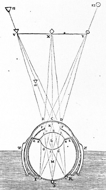
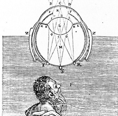
The eye as a camera (before the camera), according to Descartes.

The eye as a camera (before the camera), according to Descartes.

Vision science is not a new thing – in Europe it has been going on at least since Leonardo da Vinci tried to figure out a way to inspect the ”picture” projected inside an eye from a dead cow, hoping to see the world in the same way as a cow does. This idea was based on the same kind of misunderstanding as the diagram by Descartes that I show details of here. What misunderstanding? Well, the misunderstanding that Consciousness (of a cow or a human) is like a small person sitting somewhere inside our heads, looking at the pictures of our retinas (the ”screen” at the back wall of our eyes) like someone visiting a movie theater. The philosopher Antony Fredriksson will discuss the history of this idea in our course about moving images. Modern brain research gives ample proof that visual perception or any perception doesn’t work like that at all. In fact, there is no picture to be seen on some inner movie screen, but only a myriad of light-sensitive cells – the so called photo-receptors of the retina – each picking up a certain wavelength and intensity of light from a certain point in the environment, then sending it upwards through the numerous crossroads of the visual pathway. Somewhere – but definitely not in one place only – all these isolated signals are finally ”computed” together to form a part of our current field of consciousness. In this way, the digital camera with its receptive screen of isolated sensors is a better metaphor for vision than the traditional analogue camera.
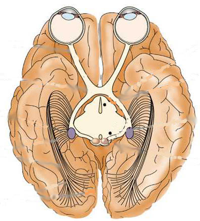
Modern picture of the basic visual pathway. The blue patches at the center are the LGNs (Lateral Geniculate Nucleae)

Modern picture of the basic visual pathway. The blue patches at the center are the LGNs (Lateral Geniculate Nucleae)

Most people know that we have two kinds of photo-receptors in the retina – rods and cones, rods being responsible for night-time vision and cones for color perception. What happens further along the road is less common knowledge. All the different signals from the photo-receptors are ”cabled” through the two optical nerves that meet at the first crossroad – called the ”optical kiasm”. That is what you see in upper half of this picture. Then the signals from the left side of the field of vision (of both eyes) are cabled to the right side of the brain, and vice versa. This happens in the ”cables” known as the visual tracts. The signals arrive at the small nucleae with the difficult name – the Lateral Geniculate Nucleus, or LGN. They are located deep inside the brain, within each side of the central region called Thalamus. The LGNs consist of at least two types of cellular layers, responsible for transmitting different kinds of information to different parts of the cortex (the cortex is the outer, ”wrinkled” parts of the brain). In this way, high-contrast information and most color information is sent through the parvo-cellular layers, while low-contrast information about movements is sent through the magno-cellular layers. It is believed that some problems with reading and writing could be due to deficiencies of the magno-cellular system.

In the lower half of the picture above, it is shown how numerous axons forward most information to the main visual center of the cortex, which is located at the very back of your head, in the region that we call the occipital lobe. And this is where we were in the course this week, learning how different cells at this ”station nr 1” (or V1) of the visual cortex are responsible for decoding specific kinds of information, for example the orientation and position of edges.
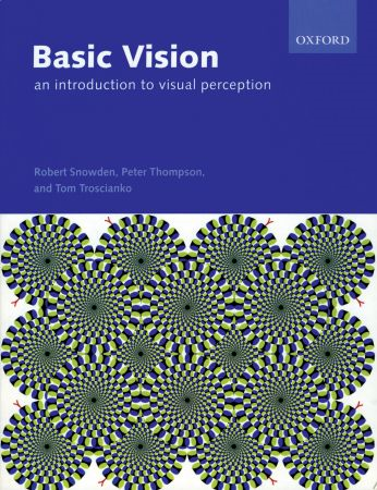
Akiyoshi Kitaokas "ormar".

Basic Vision, 2006 edition with Akiyoshi Kitaokas "snakes".

Probably our students are already busily reading the chapters for next week’s lecture, dealing with depth perception, color perception, and movement. Then, we will learn more about other visual cortex regions apart from V1 – Vision science discovers new regions all the time! Of special importance are V4 and V8 (centers for color perception) and V5 (movement perception). Look at this cover from the 2006 edition of Basic Vision. It is a popular perceptual illusion that was created by the Japanese artist and researcher Akiyoshi Kitaoka. It is basically a pattern of striped circles-within-circles, but the illusion that they are moving and the addition av small red tongues makes it easy to interpret them as snakes. Why do they seem to move? It is actually because of the division of different signals at LGN and the fact that green-black and blue-white contrast is higher than blue-black or green-white contrast. Still unclear? The exact answer is in Chapter six in Basic Vision, and in the next post on this blog.

Länkning pågår till intressant.se

Perception, Imagology and Visualization this year

Professor Anthony Johnson

Professor Anthony Johnson

After the student’s texts last week, I will now publish some posts in English, due to our current courses in English and all the positive interest that we receive from exchange students!

On this Thursday two of our courses in Visual Studies will start, and both are given in English. The first one is simply called Image Perception and Cognition, and it explains the basics in current neurological and psychological knowledge about the human visual system. You will learn some anatomy of the brain and the mechanisms at different stages in its processing of visual information. It will be a mind-boggling and exciting journey, all the way from the ”simple” registration of light at the retina, up to the cognitive job of consciously interpreting what we see!

The second course to start this Thursday is very different from the first one, so this way you really have a chance to get a clue of how broad and open the field of Visual studies really is! It is a course in arts and literature, given by our professor in English language and literature, Anthony Johnson, whom you see a picture of here. He is a legendary lecturer, and a jazz musician too.

The subject of his course is not so far away from psychological concerns as one might believe, because it is really about Imagology – a research field created by scholars who want to understand the mental images we form, for example when we read a certain text. In some respect, imagology was founded almost 100 years ago, when the American journalist and scholar Walter Lippmann developed his theory on the stereotypes we use when we describe members of certain nations or groups.

Anthony’s course is called Cultural Imagology – An introduction, and because it is about cultural imagology, he will not only talk about national and social stereotypes but also about the way people conceive of literature, geography and music. In the first lecture this Thursday, he will however begin with defining what an image is, or could be.

For those of you registered in the Image Perception and Cognition course, I will give an introduction on this Thursday, September 5th at 2 PM, in the Arken building of human sciences and languages here in Turku, room E201 (Camera Obscura). Then there is a one hour pause, and at 4 PM Anthony Johnson’s lecture starts in the same room. It will last for two hours.

Maybe you are a biologist or a student of computer systems who is also interested in art and literature, or maybe you are a student of literature who wants to know how the brain works? Then you are welcome to attend both courses!

Screen-Ddump from looking at tissue layers in BioImageXD.

Screen-dump from looking at tissue layers in BioImageXD.

In October and November I will myself give a course on Comics together with Folkloristics and Sociology. Further information in English is here. At the end of January a fourth Visual Studies course in English starts. Its name is Visuality and Visualization of Information.

This course will really give rich opportunities of combining arts with knowledge of the brain, because it is about why certain design strategies are better than others. We will use a textbook by the American psychologist Colin Ware, who is currently one of the internationally most acclaimed scientists in the expanding field Visualization.

This is a field in which neurological research, which may seem technical and abstract to many, is really put to practise in the form of effective strategies for visualization. It could be of use for marketers and web designers, but not the least for students and scholars who want to communicate research visually. There is a joint Master’s program in Bioimaging between Åbo Akademi University and Turku University, and many of our perception students have been from there. The Turku Bioimaging center (se link at top right here) has generously given all of us an opportunity to see how modern visualization works. They have created the free image processing tool BioImageXD that anyone can download at the BioImageXD page.

After downloading and opening the program, you can experiment yourself with multi-layer microscope images that you download as a free sample package at the SourceForge download page (see links at the bottom of the BioImage XD download page). The image that I show here is from a session that me and one of my students had with the program. We noticed that the possibility of generating views in 3D really added a lot to the precision of the visualization. In the Visuality and Visualization of information course we will study both simple and complex visualizations, ranging from comic books to medicine and physics, and at the end you will have the opportunity to realize a new visualization yourself.

For more inspiration, see:

”Genes to Cognition” page with an interactive 3D brain (very good for training at the Image Perception and Cognition course)

A short text defining Imagology

A Vimeo lecture on visualization by Colin Ware

/

länkning pågår till intressant.se